
Isolation of Bread Mold
To isolate different types of molds (fungi) that grow on bread To make pure cultures of different bread molds
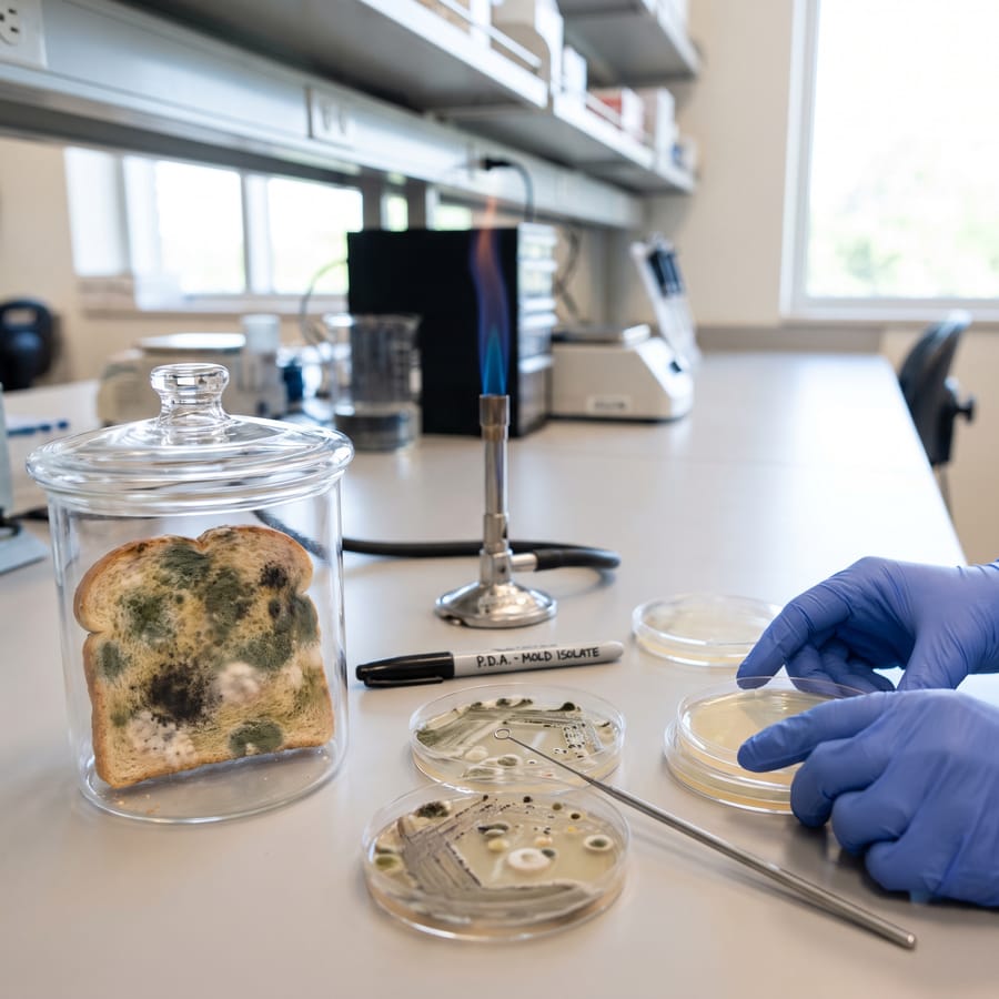
Theory & Background
Have you ever wondered about this fascinating scientific process? When bread gets old it stales or mold. You may have seen that bread molds of different colors like green, black, blue and yellow grow as cottony masses on bread surface. Different colored colonies represent different types of bread molds. Usually bread molds grow faster in damp, dark and warm conditions. Using fungal isolation techniques as described in following procedure you can prepare pure cultures of these fungi for further observations. A pure culture contains just one species of microorganism. In Microbiology pure cultures are very important in identification procedures.
Required Materials
- Sterile Potato dextrose agar medium
- Sterile Petri dishes
- Inoculation needle
- 2 Bunsen burners
- A large beaker and a glass lid
- A piece of bread
Estimated Time
2 weeks
Step-By-Step Procedure
Take a piece of bread and wet it by sprinkling water on it.
Put the bread in a beaker and cover with a glass lid/polythene.
Keep the set up 3-5 days for incubation.
When you can observe fungal masses on the bread surface, take it out and observe carefully.
Record the different colors that you can see.
Using the inoculation needle scrape a fungal colony of one color (e.g. Black colored area). Do this under aseptic conditions. Figure 1.0
Inoculate the scraped fungal mass into a potato dextrose agar (PDA) plate by streaking an “E” shape as shown in following figure. Figure 2.0
Repeat steps 6 and 7 with other colored areas (i.e. yellow, green, blue).
Label the plates and incubate for 3-5 days at 25oC.
After the incubation period you will observe well separated colonies along streak lines of PDA plates. Scrape a little from a well separated colony and place the scrape in the centre of another PDA plate Figure 3.0 Figure 4.0
Repeat the step 1 of section B with all the plate that you prepared under section A.
Incubate the plates for 3-5 days at 25oC.
Experiment Tips
- Before you proceed into procedure in section B you should get well separated colonies along streak lines of PDA plates which prepared under section A. If not, repeat the “E” streak method several times until you see separated colonies. Figure 5.0
Observation
You will observe pure colonies in final plates as in figure 6.0. Figure 6.0
Result & Conclusion
You can prepare wet mounts with isolated colonies to observe them under light microscope. You will see that morphological characters (sporangia, spores etc) of different colored colonies are different form each other. That is because these colonies of different colors are made of different species of fungi. Colony characteristics like the color, shape and appearance are very important in preliminary identification of fungi. If you get a large number of colonies in one plate, it is practically impossible to prepare wet mounts and observing them under microscope to see whether they are different species or same species. Therefore at first we look at colony characters. If the colony appearance is similar, most likely they are of same organism.